- Your cart is empty
- Continue Shopping

Product
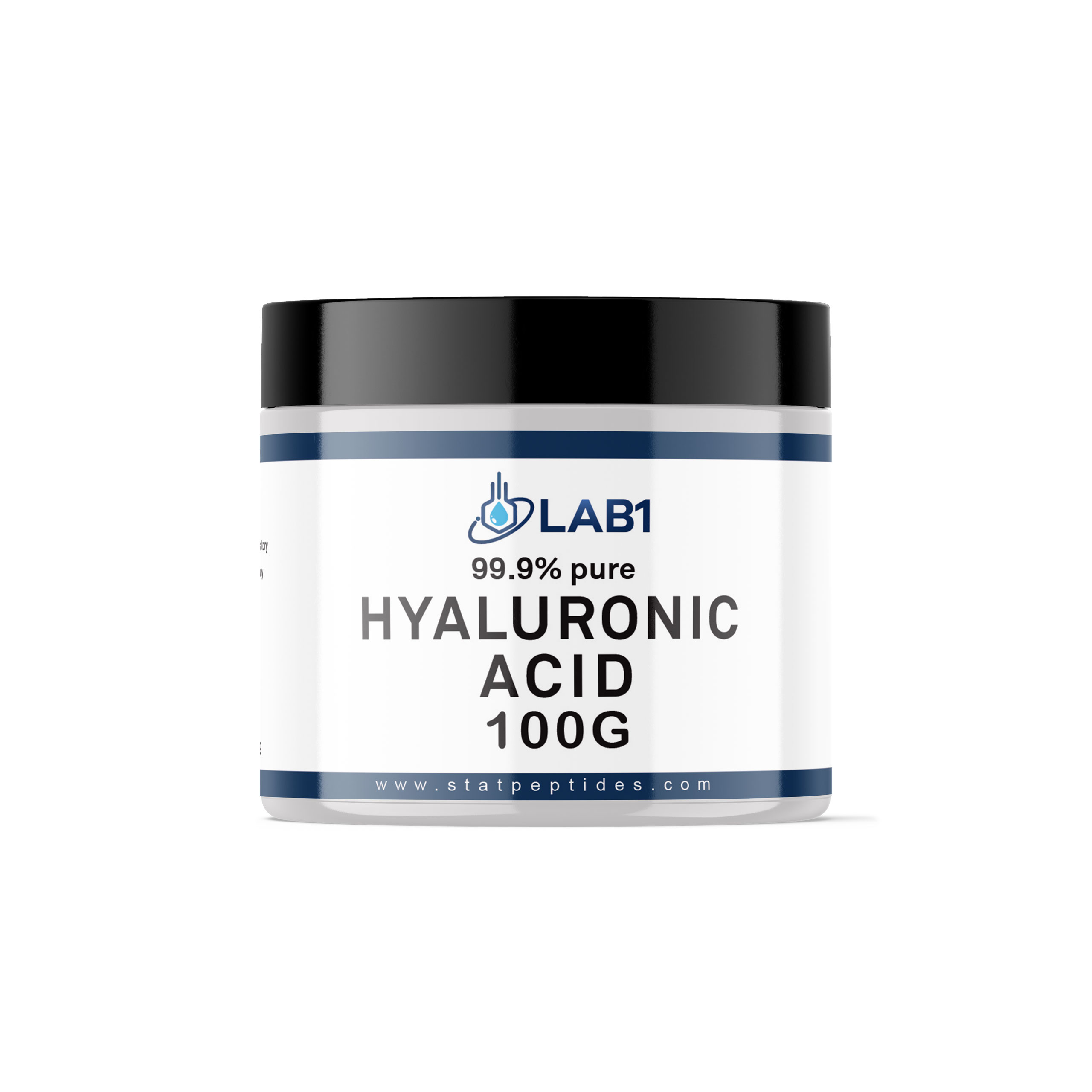
Hyaluronic Acid 100G Powder CAS 9004-61-9
$20.00
1 in stock


1.Introduction
Hyaluronic Acid (HA), also called hyaluronan, is a very important sugar-like molecule that naturally exists in our body (Necas et al., 2008). It was first discovered in 1934 by two scientists, Karl Meyer and John Palmer, while they were studying the jelly-like substance inside a cow’s eye, known as the vitreous humor. They used a chemical method to make this slippery material come out of the solution, which was the first time that HA was isolated. What makes this large molecule special is its amazing ability to pull in and hold a lot of water—working like a natural lubricant and shock absorber (Fraser et al., 1997). Because of this, HA is found in parts of the body that need moisture and cushioning, like the skin, joints, and eyes. It plays a key role in keeping tissues healthy and helping them move smoothly (Fallacara et al., 2018). Hyaluronic acid (HA) plays a scientifically proven role in boosting collagen production and supporting fibroblast activity, which are both essential for skin repair, elasticity, and overall tissue health (Kendall and Feghali-Bostwick 2014), and it is also scientifically proven that HAis present in large quantities in the embryonic stage and in newborns (Trivedi et al.,1993).
Hyaluronic acid, or HA, is a special sugar molecule that carries a tiny negative charge and does not contain sulphate. It’s made of two smaller sugar parts that repeat over and over, like beads on a long necklace. These parts are called D-glucuronic acid and N-acetyl-D-glucosamine, and these are always arranged in the same way, whether HA comes from our bodies or is made by bacteria. What makes HA truly amazing is its water loving property. Because of its water-attracting groups, it can soak up water like a sponge—up to 1,000 times of its own weight (Kielbowicz et al., 2024). This gives HA a jelly-like texture that’s thick when still, but becomes smooth and slippery when moved. That’s why it works so well inside our joints, helping them move easily, and in our skin, keeping it soft, full, and hydrated (Dubrovin et al., 2023).
Hyaluronic acid (HA) works like a flexible switch in the body, and its job depends mostly on its size. When HA is large called High-Molecular-Weight HA (HMW-HA) it forms a thick, watery layer that helps build the structure around cells, known as the extracellular matrix (ECM). This layer gives tissues strength, keeps them firm, and helps joints move smoothly (Jędrak & Dolińska, 2024). In this form, HA acts quietly, filling space and absorbing shocks. But when enzymes or stress break HA into smaller pieces called Low-Molecular-Weight HA (LMW-HA) it becomes active and starts sending signals to cells (Di Mola et al., 2022). These signals go through special cell receptors like CD44 and RHAMM. For example, large HA binding to CD44 can reduce inflammation, while small HA pieces binding to the same receptors or to others like TLR2/4 can increase inflammation and help form new blood vessels, which is important for healing wounds (Jędrak & Dolińska, 2024). So, HA works in two ways: the large form keeps tissues stable and healthy, while the small form helps cells grow, move, and respond during tissue repair.
Hyaluronic acid (HA) plays an important role in tissue engineering, especially in the process of decellularization and recellularization (Neishabouri et al., 2022). Decellularization means removing all the cells and genetic material from natural tissue to create a clean, three-dimensional scaffold. This scaffold keeps the structure of the extracellular matrix (ECM), which is important for supporting new cells (Chen et al., 2023). However, the strong steps used during decellularization can sometimes lower the amount of HA in the scaffold, which can affect its natural properties (Neishabouri et al., 2022). To solve this problem, HA is often added back into the scaffold or chemically changed to improve cellularization which is the process of adding new cells. A HA rich scaffold is very helpful because it copies the natural ECM, improves compatibility with cells, and gives the right signals to guide them. The added HA helps new cells stick to the scaffold, grow, and change into the needed types—like stem cells turning into cartilage or skin—which is essential for rebuilding healthy tissues (Rajabi et al., 2022).
Hyaluronic acid (HA) has high water holding capacity due to which it works like a smooth lubricant. Because of this, it is used in many areas of medicine and industry. In skin care, HA keep the skin moist and stretchy, so it often used in anti-aging products and cosmetic fillers (Jędrak & Dolińska, 2024). In bone and joint care, HA injections are used for better joint movement and pain reduction, especially in people with osteoarthritis (Kielbowicz et al., 2024). Due to its transparent and protective nature, it is used to treat dry eyes in eyes surgeries. (Di Mola et al., 2022). In tissue repair and regenerative medicine, HA’s gel-like form acts like soft support for growing new tissues like skin and cartilage. HA also plays a role in cancer. It can help cancer cells to grow by connecting with a receptor called CD44. But scientists also use this same connection to deliver cancer medicine. They make HA-coated particles that carry chemotherapy drugs directly to tumor cells, helping protect healthy cells from damage (Pashkina et al., 2024).
Hyaluronic Acid (HA) has come a long way from being taken from animal tissues to now being made using bacteria. It has special properties like holding water and acting like a soft gel, which help keep tissues strong and hydrated (Jędrak & Dolińska, 2024). HA also connects with cell receptors like CD44, making it useful in tissue repair. It supports new cell growth (cellularization) and works well in scaffolds made by removing old cells (decellularization) (Rajabi et al., 2022). Because of this, HA is now used in many areas—like skin care, joint treatments, and targeted drug delivery systems. One of its most important uses is in cancer treatment, where HA helps guide medicine directly to tumor cells using the CD44 receptor, helping protect healthy cells (Pashkina et al., 2024).
What is Hyaluronic acid
Research shows that hyaluronic acid (HA) was first discovered in 1934 by scientists Karl Meyer and John Palmer while studying the clear gel inside cow eyes, which they named after the Greek word hyalos, meaning gloss (Meyer & Palmer, 1934). Further studies reveal that while HA was originally extracted from animal tissues like rooster combs, modern production now relies on bacterial fermentation, a biotechnology method that yields purer, non-animal-derived HA and reduces the risk of immune reactions (Kielbowicz et al., 2024). Research also shows that HA is made of repeating sugar units—D-glucuronic acid and N-acetyl-D-glucosamine—that form a long, unbranched chain, giving it unique biological properties. Most importantly, research confirms that HA has an extraordinary ability to hold water—up to 1,000 times its own weight, that’s why it plays such a vital role in keeping our skin hydrated, our joints lubricated, and our eyes moist (HTL Biotechnology, 2025).
Chemical structure and Physiochemical properties
Consider hyaluronic acid (HA) as a long, silky thread made of sugar units, gently weaving through our body’s tissues. Research shows that HA is a special type of natural molecule called a glycosaminoglycan—a long, unbranched sugar chain with a high molecular weight (Fallacara et al., 2018). Its basic structure is like a repeating pattern, where each unit has two sugar-like building blocks: one called D-glucuronic acid and the other N-acetyl-D-glucosamine. These two sugars are tightly linked together in an alternating fashion, like beads connected by two types of bonds—β−1,4 and β−1,3 glycosidic linkages—which help form a smooth, extended chain (Necas et al., 2008). The number of these repeating units, shown as ‘n’ in its chemical formula (C₁₄H₂₁NO₁₁) n, can vary a lot. The longer the chain, the heavier the molecule—and this size difference directly affects how HA behaves in the body, from hydrating tissues to supporting healing and elasticity (Fallacara et al., 2018).
- Polyanionic nature facilitates binding to water molecules, maintaining tissue hydration.
- High molecular weight varies widely depending on tissue source and physiological conditions.
- Non-sulfated structure distinguishes it from other glycosaminoglycans.
The physicochemical properties of HA are largely dictated by its molecular weight, which can range from a few kilodaltons (kDa) to several million Daltons (MDa). High-Molecular-Weight HA (HMW-HA, >1,000 kDa) provides structural integrity and viscoelasticity. Imagine hyaluronic acid (HA) as a sponge-like molecule with a clever design—its structure is packed with special groups called hydroxyl (−OH) and carboxyl (−COOH), which love water (Necas et al., 2008). Because of this, research shows that HA is incredibly hydrophilic, meaning it can soak up water like a champ—just one gram of it can hold up to six liters. When HA absorbs water, it transforms into a thick, stretchy gel that forms a soft, three-dimensional network known as a viscoelastic solution (Kielbowicz et al., 2024). This gel-like texture is what makes HA so useful in the body, it cushions our joints like a shock absorber, keeps our skin plump and hydrated, and fills space between cells to maintain tissue structure. HA is naturally safe and gentle because its basic structure is the same in all vertebrates, which means it rarely causes allergic reactions and is widely used in both medicine and skincare (Dubrovin et al., 2023).
Importance of Hyaluronic acid
1.HA in Infancy: A Biomarker and a Healing Agent
Research shows that hyaluronic acid (HA) plays a powerful role in helping newborn babies grow and stay healthy. Right after birth, babies have very high levels of HA in their blood, much more than older children or adults. That’s because HA is a key part of the body’s building blocks, especially in fast-growing areas like the liver and soft tissues. It’s made and cleared quickly by special cells in the liver, and scientists have found that these high levels are not just normal, they’re helpful.
In fact, research shows that HA can act like an early warning signal for liver problems in babies, such as biliary atresia or alpha-1 antitrypsin deficiency, often showing up before regular liver tests detect anything (Trivedi et al., 1993). But HA doesn’t stop there. It also has soothing powers—it keeps tissues moist, reduces inflammation, and helps repair damage. That’s why doctors sometimes use HA in a mist (nebulized form) to help babies recover from breathing problems, as it hydrates the airways and supports healing (Licari et al., 2015). So, from liver health to lung care, HA quietly supports newborns in their earliest and most delicate days.
- Role of HA in boosting collagen:
Research shows that hyaluronic acid does not turn into collagen instead, it creates the perfect environment for collagen production. HA keeps the skin’s surroundings hydrated, cushioned, and healthy, which allows fibroblasts-the skin’s natural repair cell- to work at their best. Think of fibroblasts as skin’s construction crew. Their job is to build and repair collagen, keeping skin firm, smooth and supported. When these cells are surrounded by moisture-rich HA, they stay active and can produce collagen more efficiently. This is why HA is often associated with skin repair and antiaging benefits. It does not become collagen it helps your cells make it better. Supported by research: HA has been shown to stimulate fibroblast activity and enhance collagen synthesis by improving the cellular environment. (Papakonstantinou et al.,2012; Kendall&Feghali-Bostwick,2014)
3.Biological Roles and Mechanisms of Hyaluronic Acid
Research shows that hyaluronic acid (HA) is a shapeshifter in the body—its role depends entirely on its size, or molecular weight (MW). When HA is large and heavy, it acts like a builder and protector. But when it breaks into smaller pieces, it turns into a messenger that signals damage and calls for repair. (Fallacara et al., 2018).
- High Molecular Weight HA (HMW-HA): The Builder
This form of HA, with a molecular weight over 1 million Daltons, is found in healthy places like the jelly of the eye and the fluid in our joints. It’s thick, stretchy, and holds water like a sponge, it cushions joints and keeps tissues firm and elastic (Necas et al., 2008). But HMW-HA does more than just support—it also plays role in protection by forming a gentle barrier that blocks harmful immune cells from entering healthy tissue, keeping inflammation in check and helping cells stay calm and balanced (Fallacara et al., 2018)
- Low Molecular Weight HA (LMW-HA): The Messenger
Research show that when the body gets hurt—through injury, infection, or stress—HMW-HA is chopped into smaller pieces by enzymes called hyaluronidases (Necas et al., 2008). These fragments, usually under 500,000 Daltons, act like alarm bells. Scientists call them DAMPs—Danger-Associated Molecular Patterns—because they alert the immune system that something’s wrong (Fallacara et al., 2018). LMW-HA doesn’t just shout for help, it sparks inflammation, attracts immune cells, and helps build new blood vessels, all of which are vital for healing wounds (Fallacara et al., 2018).
4.Interaction with Cell Receptors
HA delivers its messages by connecting with special receptors on cell surfaces. These receptors “read” the size of the HA and respond accordingly.
- CD44 (The Primary HA Receptor)
CD44 is the most common HA receptor in the body and is found on nearly every type of cell, including those that line our organs (epithelial cells), blood vessels (endothelial cells), and those that defend us from disease (immune cells) (Necas et al., 2008). This receptor plays a key role in how cells stay connected to their surroundings and how they move when needed. When HA is in its large, high-molecular-weight form (HMW-HA), it binds to CD44 and helps cells stick to the tissue framework, keeping everything stable and well-organized—like bricks held together by mortar. But when HA breaks down into smaller fragments (low-molecular-weight HA or LMW-HA), the story changes. These tiny pieces still bind to CD44, but instead of holding things in place, they send signals inside the cell that trigger growth, survival, and movement. This signaling is especially active during healing—but research shows that cancer cells can hijack this same pathway to spread and invade other tissues (Fallacara et al., 2018). So, depending on its size, HA’s interaction with CD44 can either build and protect or activate and mobilize, making it a powerful player in both health and disease.
- RHAMM: (Receptor for Hyaluronan-Mediated Motility)
RHAMM is found on fast-moving cells like immune cells and those involved in wound healing. Research shows that RHAMM is especially important for cells that need to move quickly—like immune cells rushing to fight infection, or cells involved in early development and wound healing (Jian et al., 2020). RHAMM works by connecting with hyaluronic acid (HA), and when HA binds to it, it gives cells the signal to start moving. This movement, known as cell migration, is crucial during tissue remodeling—when the body needs to reorganize and send cells into damaged areas to fix and rebuild (Litwiniuk et al., 2016). Without RHAMM guiding this process, healing would be slower and less efficient. So, whether it’s sealing a wound or shaping a developing organ, RHAMM and HA work together like a GPS and fuel system, helping cells find their way and keep going until the job is done (Jian et al., 2020).
- Toll like Receptors:(TLR2 and TLR4)
When the body senses danger—like an injury, infection, or stress—it needs a fast and powerful way to call for help. That’s where the tiniest pieces of hyaluronic acid (HA), called oligosaccharides, step in. Research shows that these small HA fragments act like emergency signals by directly connecting with special sensors on immune cells known as Toll-like receptors, specifically TLR2 and TLR4, which are mostly found on cells like macrophages—the body’s frontline defenders (Fallacara et al., 2018). Once these fragments bind to the receptors, they flip a molecular switch that sets off the body’s built-in alarm system. This triggers the rapid release of pro-inflammatory messengers called cytokines and chemokines, which rush to the scene and amplify the immune response (Necas et al., 2008). It sounds like a siren that alerts the immune system to act—fast—so healing can begin right away.
5. Role of HA in extracellular matrix
Research shows that hyaluronic acid (HA) plays a starring role in the body’s support system, known as the extracellular matrix (ECM)a complex network that surrounds and holds cells together in connective tissues (Fallacara et al., 2018). What makes HA so unique is that, unlike other glycosaminoglycans (GAGs), it doesn’t attach to a protein core. Instead, it floats freely as a long, sugar-based polymer, earning its place as the only non-sulfated GAG in the ECM (Necas et al., 2008). Its superpower lies in its love for water—HA is extremely hydrophilic, meaning it can soak up and hold huge amounts of moisture. This creates a soft, gel-like cushion that fills space, resists pressure, and keeps tissues plump and hydrated (Jian et al., 2020).
HA doesn’t work alone, in its high-molecular-weight form (HMW-HA), it acts like a scaffold, helping other important ECM molecules like Versican and Aggrecan come together to form large, stable structures. It also helps organize fibrous proteins like fibronectin and Type III collagen, which are key for building tissues and healing wounds with minimal scarring (Necas et al., 2008). Because HA is so important, the body carefully controls how much it is made and broken down. Changes in its size and concentration send signals to cells, guiding how they move, grow, and transform—making HA a master coordinator of tissue health and repair (Fallacara et al., 2018).
6. Hyaluronic Acid in Decellularization and Recellularization
- Decellularization and Tissue Scaffold:
Research shows that regenerative medicine often uses special frameworks called scaffolds to help the body grow new tissue. These scaffolds are designed to mimic the body’s natural extracellular matrix (ECM)the supportive network that surrounds cells (An et al., 2021). To create them, scientists use a process called decellularization, which carefully removes all living cells and genetic material from tissues or organs. What’s left is a clean, natural structure that keeps the original shape, strength, and chemical makeup of the tissue (Lin et al., 2015; Jayasree et al., 2023).
- Role in Decellularization of Scaffolds:
Research shows that hyaluronic acid (HA) is a vital part of the native ECM, so adding it back into decellularized scaffolds makes them more effective (Jayasree et al., 2023). For example, scaffolds made from pig peritoneum for skin repair are often coated with HA. This helps them hold onto important growth signals like basic fibroblast growth factor (bFGF) and epidermal growth factor (EGF), which guide cells to grow and heal (Lin et al., 2015). In cartilage repair, even when strong chemicals like hydrochloric acid are used to remove cells, HA’s gentle and biocompatible nature helps restore the scaffold’s healing potential during the next step—recellularization (Nürnberger et al., 2021).
- Role in Recellularization:
Research shows that recellularization, is the process of adding new cells—often from the patient—onto the scaffold to rebuild tissue (An et al., 2021). HA-based materials, especially soft gels called hydrogels, are ideal for this because they resemble the body’s natural ECM and hold lots of water (Chang, 2025). These HA scaffolds create a friendly space where cells can stick, grow, and move. The presence of HA also helps guide cell behavior, encouraging important cells like fibroblasts and endothelial cells to multiply and travel to the damaged area (Jayasree et al., 2023). Furthermore, research shows that HA can calm inflammation and help stem cells survive and settle in, making it a powerful tool in advanced cell therapies (Chang, 2025).
7. Research Applications in medicine and industry
Due to its multifunctional properties, HA has a broad spectrum of applications across different fields:
- Osteoarthritis and Joint Health
Research shows that intra-articular HA injections are widely used for osteoarthritis treatment, serving as a therapy known as Visco supplementation to restore joint mechanics. These injections provide lubrication to the synovial fluid, reducing friction between the articulating cartilage surfaces (Bowman et al., 2018). HA’s high viscosity helps protect the remaining cartilage from further mechanical damage and degradation. The therapeutic effect also involves anti-inflammatory action, inhibiting pain receptors and reducing inflammatory mediators in the joint space (Stat Pearls, 2023). The success of this treatment is often dependent on the specific molecular weight and concentration of the HA formulation used (Bowman et al., 2018).
- Dermatology and Cosmetics
Research studies show that HA formulations significantly improve skin hydration by attracting and retaining large volumes of water within the epidermis. This strong hygroscopic property helps to restore and reinforce the skin barrier function, thereby minimizing trans epidermal water loss. HA-based dermal fillers are standard cosmetic treatments, instantly correcting deep wrinkles and restoring lost facial volume due to their injectable gel-like consistency. Clinical trials consistently confirm HA’s efficacy in reducing signs of aging, contributing to improved skin elasticity and a plumper, smoother appearance (Robinson et al., 2022).
- Oral Supplementation
It is scientifically proven that oral HA supplements show potential in improving systemic skin hydration and elasticity by providing raw material for internal synthesis. The low molecular weight HA fragments are absorbed in the gastrointestinal tract and are believed to be distributed to the skin and connective tissues. Some studies also suggest systemic benefits for joint function, potentially by increasing the concentration of HA precursors in the synovial fluid. Additionally, oral intake of HA has been implicated in accelerating wound repair and promoting tissue recovery across various bodily tissues (Gao et al., 2021).
- Biomaterials and Tissue Engineering
Research shows that crosslinked HA hydrogels are highly versatile biomaterials that serve as bio-mimetic scaffolds for cartilage and bone regeneration. These scaffolds are designed to provide structural support while possessing a high-water content that closely mimics the native extracellular matrix (ECM) environment (Burla et al., 2019). The HA-based scaffolds promote cellularization by allowing cell migration, proliferation, and differentiation of seeded cells (e.g., stem cells) throughout the three-dimensional matrix (Chang, 2025). Furthermore, HA’s native components can be leveraged in decellularization protocols, where its structure in the natural ECM acts as a template before being supplemented back into the acellular scaffold to enhance tissue repair (Lin et al., 2015). The chemical versatility of HA allows researchers to tune the mechanical stiffness and degradation rate of the hydrogels to match the specific tissue being regenerated (Chang, 2025).
- Cancer Biology and Drug Delivery
Research studies suggest that HA is actively explored in cancer drug delivery due to its high affinity for the CD44 receptor, a surface marker often overexpressed on malignant tumor cells. Nanoparticles are frequently coated or conjugated with HA, leveraging this receptor-mediated targeting to deliver chemotherapeutic drugs preferentially to the tumor site (Gao et al., 2021). This targeted approach significantly enhances drug efficacy while minimizing systemic toxicity to healthy organs and tissues (Misra et al., 2015). Emerging research focuses on developing HA-based imaging agents for cancer diagnostics, utilizing the same CD44-targeting principle for non-invasive tumor visualization and early detection (Gao et al., 2021).
Conclusion
Hyaluronic acid (HA), also called hyaluronan, is a natural sugar-based molecule found all over the body. It plays a key role in how our tissues are built and how they function (Necas et al., 2008). Back in 1934, scientists Karl Meyer and John Palmer first discovered HA in the jelly-like fluid of a cow’s eye. Since then, decades of research have revealed that this once-overlooked molecule is far more than just a filler in the body—it’s now known as a powerful regulator of how cells behave. Its effects depend heavily on how much of it is present and how big its molecules are (Fallacara et al., 2018)
Chemically, HA is a type of glycosaminoglycan made up of repeating sugar units. Its formula is (C₁₄H₂₁NO₁₁)ₙ, and each unit includes two sugars: D-glucuronic acid and N-acetyl-D-glucosamine, linked together in a special pattern (Casale et al., 2016). What makes HA truly special is its extreme love for water. Research shows that just one gram of HA can hold up to six liters of water (Jian et al., 2020). This gives tissues their bounce, stretch, and ability to absorb shock—especially in places like joints, where HA acts as a natural lubricant (Litwiniuk et al., 2016).
In newborn babies, HA levels in the blood are naturally high. These elevated levels help support rapid tissue growth and development. Interestingly, they also serve as early warning signs for liver-related diseases like biliary atresia, even before standard tests show anything wrong (Trivedi et al., 1993). When it comes to healing, HA plays a major role by guiding fibroblasts—the cells that build the tissue matrix. It helps them organize collagen and fibronectin fibers, which are essential for forming new tissue during growth and wound repair (Jian et al., 2020; Necas et al., 2008).
As a key part of the extracellular matrix (ECM), HA acts like a soft scaffold that holds everything together. It helps large molecules like proteoglycans stick together and keep tissues hydrated and strong (Fallacara et al., 2018). HA also sends signals to cells through special receptors like CD44 and RHAMM. When high-molecular-weight HA binds to these receptors, it helps cells stay in place and rest. But when HA breaks into smaller pieces, it activates pathways that tell cells to grow, move, and multiply—an important feature in tissue repair and regeneration (Litwiniuk et al., 2016; Jian et al., 2020).
HA is now widely used in both medicine and industry. In the pharmaceutical world, it’s used to deliver drugs directly to target tissues—especially tumors that have lots of CD44 receptors (Fallacara et al., 2018). In the medical field, HA is used in eye surgeries, joint injections for arthritis, and even in cosmetics as a key ingredient in dermal fillers and anti-aging products because of its amazing ability to hold moisture (Casale et al., 2016).
However, natural HA isn’t perfect. One of its main drawbacks is that it breaks down quickly in the body and doesn’t stay stable for long—especially in implants or drug delivery systems (Jian et al., 2020). That’s why researchers are now working on ways to improve it. By chemically modifying HA or cross-linking its molecules, they’re creating new forms like hydrogels that last longer, degrade at controlled rates, and offer better strength. These advanced versions of HA are paving the way for the next generation of tissue engineering, regenerative therapies, and highly targeted drug delivery systems (Fallacara et al., 2018; Jian et al., 2020).
Safety, Handling, and Regulatory Perspective
Although HA is considered safe and biocompatible, appropriate laboratory practices are essential.
- Store HA powder in airtight, moisture-free containers at room temperature.
Prepare solutions under aseptic conditions to avoid microbial contamination. Store liquid formulations at refrigerated temperatures
- The U.S. Food and Drug Administration (FDA) approve HA-based dermal fillers and Visco supplementation for osteoarthritis.
- The European Medicines Agency (EMA) regulates HA in both therapeutic and cosmetic applications.
- The World Health Organization (WHO, 2021) emphasizes maintaining quality, purity, and sterility for biomaterials, particularly in joint therapies. Overall, HA remains one of the most reliable biomolecules in biomedical and cosmetic research (Stat Pearls, 2023).
Why Choose Stat Peptide for Research-Grade Hyaluronic Acid?
Stat Peptide is a trusted provider of high-quality HA specifically designed for research applications.
- Produced via advanced fermentation and multi-step purification processes.
- Research-grade purity with minimal endotoxin levels.
- Rigorous quality control including molecular weight profiling and purity confirmation.
- Certificates of Analysis (CoA) provided for transparency.
By prioritizing reproducibility and scientific rigor, Stat Peptide enables researchers to achieve consistent and validated results in their experiments.
References
- Fallacara, A., Baldini, E., Manzotti, E., & Verga, L. (2018). Hyaluronic acid in the third millennium. Polymers, 10(7), 701.
- Fraser, J. R. E., Laurent, T. C., & Laurent, U. B. G. (1997). Hyaluronan: its nature, distribution, functions and turnover. Journal of Internal Medicine, 242(1), 27–33.
- Necas, J., Bartosikova, L., Musil, J., & Kriz, P. (2008). Hyaluronic acid (hyaluronan): a review. Veterinarni Medicina, 53(8), 397–411.
- Dubrovin, E. V., Barinov, N. A., Ivanov, D. A., & Klinov, D. V. (2023). Single-molecule AFM study of hyaluronic acid softening in electrolyte solutions. Carbohydrate Polymers, 303, 120472.
- Kielbowicz, A., Ratajczak, M., Kielbowicz, Z., Ziemkiewicz, A., Szuta, S., & Bocheńska, P. (2024). Hyaluronan: Sources, structure, features and applications. Molecules, 29(3), 739.
- Di Mola, A., Landi, M. R., Massa, A., D’Amora, U., & Guarino, V. (2022). Hyaluronic acid-based wound dressing with antimicrobial properties for wound healing application. Applied Sciences, 12(6), 3091.
- Jędrak, M., & Dolińska, B. (2024). Hyaluronic acid and skin: Its role in aging and wound-healing processes. Molecules, 29(4), 281.
- Neishabouri, A., Soltani Khaboushan, A., Daghigh, F., Kajbafzadeh, A., & Majidi Zolbin, M. (2022). Decellularization in tissue engineering and regenerative medicine: Evaluation, modification, and application methods. Frontiers in Bioengineering and Biotechnology, 10, 805299.
- Rajabi, A., Farhadian, S., Khosravi, A., & Bavafa, S. (2022). In vitro study of hyaluronic acid-based scaffolds and their effect on cartilage regeneration. International Journal of Medical Laboratory, 9(1), 6-16.
- Chen, X., He, J., Zhang, M., Su, W., Wu, M., Shi, S., & Li, Y. (2023). Recent advances in fabrication of dECM-based composite materials for skin tissue engineering. Frontiers in Bioengineering and Biotechnology, 12, 1348856.
- Pashkina, E. A., Bykova, M. S., Kozlov, V. K., & Berishvili, M. N. (2024). Hyaluronic Acid-Based Drug Delivery Systems for Cancer Therapy. Cells, 14(2), 61.
- Trivedi, P., P. Cheeseman, and A. P. Mowat. 1993. “Serum hyaluronic acid in healthy infants and children and its value as a marker of progressive hepatobiliary disease starting in infancy.” Clinica Chimica Acta 215 (1): 29–39. doi:10.1016/0009-8981(93)90246-z.
- HTL Biotechnology. (2025). Decoding Hyaluronic Acid: Its Biological Function and Uses.
- Meyer, K., & Palmer, J. W. (1934). The polysaccharide of vitreous humor. The Journal of Biological Chemistry, 107(3), 629–634.
- Licari, A., De Filippo, M., Caimmi, M., Vianello, A., Vianello, S., Mappa, L., Ruggieri, M., Cazzato, S., & Marseglia, G. L. (2015). Hyaluronic Acid and upper airway inflammation in pediatric population: A systematic review. Frontiers in Pharmacology, 6, 241.
- Jian, F., Wang, Y., Zhu, Y., Li, Q., Sun, Z., & Chen, H. (2020). Advances in the chemical modification of hyaluronic acid and its biological applications. Carbohydrate Polymers, 240, 116345. https://doi.org/10.1016/j.carbpol.2020.116345
- Gao, F., Cao, M., Lv, R., Zhang, C., & Zhao, P. (2021). Oral administration of hyaluronan improves skin hydration and elasticity. Skin Research and Technology, 27(4), 499–507.
- Bowman, S., Awad, M. E., Hamrick, M. W., Hunter, M., & Fulzele, S. (2018). Recent advances in hyaluronic acid-based therapy for osteoarthritis. Frontiers in Veterinary Science, 5, 153.
- Robinson, J. J., et al. (2022). Efficacy of topical hyaluronic acid in improving skin hydration: A randomized controlled trial. Journal of Cosmetic Dermatology, 21(2), 421–429.
- Papakonstantinou, E., Roth, M., Karakilulakis, G (2012). Hyaluronic acid: Akey molecule in skin aging. Dermato-Endocrinology,4(3),253-258
- Kendall, R.T., & Feghali-Bostwick, C. (2014). Fibroblasts in fibrosis: Novel roles and mediators. Fronteirs in Pharmacology, 5, 123
Safety Data Sheet will be available soon.
| Weight | 2.35 oz |
|---|---|
| Dimensions | 2.84 × 2.84 × 2.88 in |